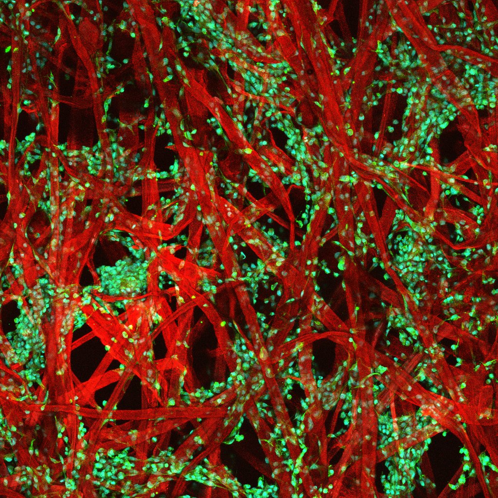
Description: Understanding how cells are influenced by their environment is crucial for developing new therapies for cancer. But common methods of growing and studying cells in the lab, such as two-dimensional petri dishes or animals, fall short of replicating these complex interactions. Tissue engineering offers a better way. In the McGuigan lab, we are growing cells on scaffolds made of cellulose — a key component of paper — and rolling them up like Swiss roll cakes. Called TRACER, these layered, three-dimensional structures offer a new way of studying cancer by reproducing many of the features of real tumors. For example, in TRACER there is formation of oxygen gradients like the one found in tumors. Using TRACER, we can then study the impact of oxygen level on cancer cell behaviour (proliferation, drug resistance, etc..). Understanding the regulation of cancer cells by the microenvironment is critical to develop new therapies. More broadly, this research has application in other fields such as regenerative medicine, where knowing how to modify cell behaviour with external clues is essential.
The technology we developed to understand how cellular behaviour is regulated by its microenvironment was inspired by cake. Answering the question, however, is not a piece of cake.
Why did you conduct this research? My goal is to understand how the level of oxygen influences the way cells communicate as in several pathologies (cancer, fibrosis, rheumatoid arthritis) the level of oxygen in the tissues is dysregulated. Using TRACER, I can compare how cells behave at the outside of the “Swiss roll” where there is lot of oxygen, versus the cells located at the core of the “Swiss roll” where there is less oxygen. Understanding how oxygen modify cell behaviour could lead to the discovery of new targets for future therapies.
Technique: To obtain this image we used three different labelling that color specifically : the paper fibers (in red), the cells (in green) and the cells nucleus (in cyan). After labelling, the piece of paper was sandwiched between two pieces of glass to maintain it flat and to make it compatible with the use of microscopy. We then used a special microscope called confocal microscope that produce really contrasted images. This microscope take one picture per color, then using a computer and a special software we just merge the 3 colors to obtain the final image.
Acknowledgements: Dr. Latour is supported by a post-doctoral fellowship from the University of Toronto’s Medicine by Design initiative, which receives funding from the Canada First Research Excellence Fund (CFREF).

