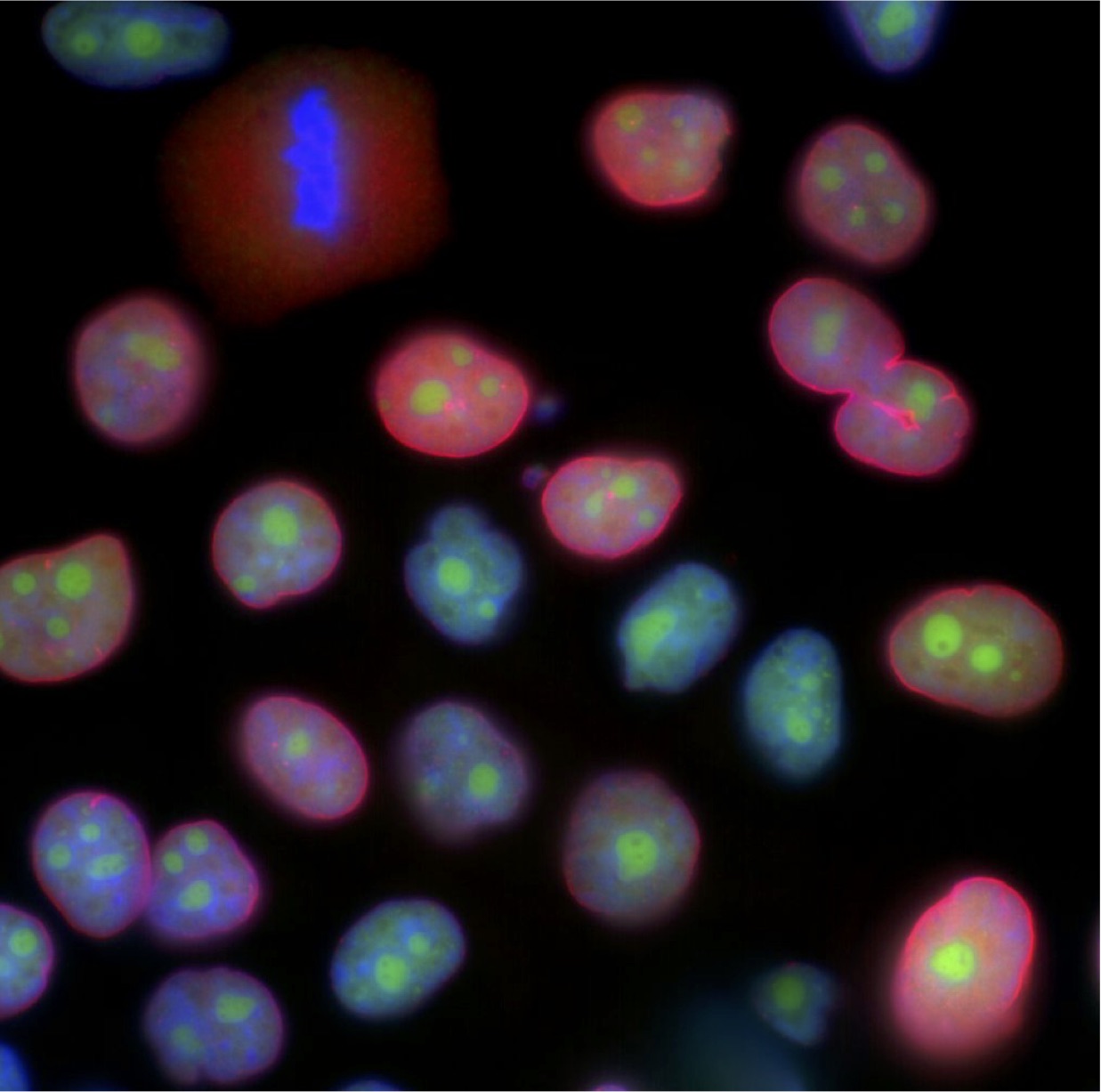
Description: I’m a cell biologist, interested in how our cells respond to different kinds of DNA damage, including radiation exposure or chemotherapy. This means I spend a lot of time looking at both healthy and damaged cells under a microscope. I use fluorescently labelled antibodies to visualize different cellular structures in different colours. In this picture, DNA is in blue, active gene transcription is in green, and the cell’s nuclear envelope is in red. Almost all the cells in this picture look healthy and intact, with a red nuclear envelope encircling their blue DNA, with pockets of transcription occuring in green. But in the top-left corner, I caught a cell going through mitosis. Its nuclear envelope has broken down, leaving a cloud of red particles. Its chromosomes have condensed into the blue ball in the middle of the cloud, replicated and ready to separate into two daughter cells. I caught this cell at exactly the right moment, with its DNA completely exposed. In another hour, it would have finished separating the ball of DNA into two identical, complete sets of chromosomes. It would have re-formed two daughter nuclei, and would have looked like every other cell in the picture.
Why did you conduct this research? I want to know whether our cells respond differently to different kinds of DNA damage. The ways in which cells react to DNA damage can influence their development into cancer, since cells that don’t repair damage are more likely to accumulate cancer-causing mutations. Investigating responses to specific types of DNA damage will help us understand more about the process that transforms a healthy cell into a cancerous one. Taking pictures of healthy cells and damaged cells is one of the many ways I can demonstrate such a response.
Technique: These cells are from a cultured human cell line, called MCF10A. They were originally derived from human breast epithelial cells, but they’ve been transformed into an immortal cell line, to be used exclusively for research. I used immunofluorescent antibodies to mark each of the three cellular structures in the image (DNA, active transcription, and the nuclear envelope), and then used a fluorescent microscope to take the picture.
Acknowledgements: The Canadian Institutes of Health Research, the University Health Network, and the University of Toronto have all funded my research. I would also like to acknowledge the Harding Lab.

