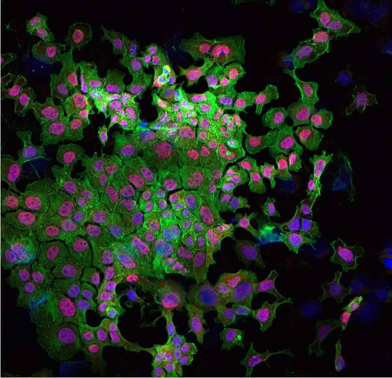
Description: This is a confocal microscopy image of MLE12 mouse lung epithelial cells, which were stained with several fluorescent dyes to visualize the activity of signalling molecules Yap and Taz (in red). It was taken at the Lund Bioimaging Center at Lund University in Sweden while I was a member of a summer research abroad project in the Lung Bioengineering and Regeneration lab. The image was captured after the cells had been treated for 6 hours with Transforming Growth Factor (TGF-β), a key signalling player in a fatal lung disease called Idiopathic Pulmonary Fibrosis (IPF). TGF-β was used to mimic fibrotic changes in the MLE12 cells and investigate whether Yap/Taz complexes subsequently move into the nuclei of the cells (in blue), where they carry out their effects on gene expression in IPF. As seen by the overlap of red and blue staining in the nucleus, increased nuclear localization of Yap/Taz was observed after 6 hours of TGF-β treatment. This provides evidence that the effects of TGF-β signalling in IPF may be mediated by Yap/Taz. TGF-β can thus be used to induce fibrosis before targeting Yap/Taz and observing changes in fibrotic expression, as was done in later parts of this research project.
Why did you conduct this research? Although TGF-β signalling is known to induce fibrotic changes in IPF, it was unclear whether its effects were mediated by Yap/Taz activity in the nucleus. This experiment was the first step in confirming TGF-β-induced Yap/Taz activation before inhibiting the complex with a drug called Verteporfin. Verteporfin reduced fibrotic expression, but the effects of TGF-β continued to be observed. Although this points to additional TGF-β signalling mediators in IPF, our ability to target Yap/Taz is promising for continuing studies that assess VP in IPF treatment. Current treatment options are extremely limited and none effectively cure IPF.
Technique: Several staining dyes were used to visualize components of the MLE12 cells under a Nikon A1 + confocal microscope at 20x magnification. A blue-fluorescent DNA stain called 4′,6-diamidino-2-phenylindole (DAPI) was used to outline the nuclei of the cells. The cytoskeleton is a structural frame that supports the cell’s shape and was stained with an actin filament probe called Phalloidin that was attached to a green-fluorescent dye. Lastly, Yap/Taz was stained using a Yap/Taz secondary antibody protein attached to a red fluorescent compound. The composite image of all 3 stains was created using ImageJ.
Acknowledgements: I would like to thank Dr. Darcy Wagner and Hani Alsafadi for generously sharing their time and knowledge with me throughout this project. I would also like to acknowledge that this image was taken at the Lund University Bioimaging Center.

