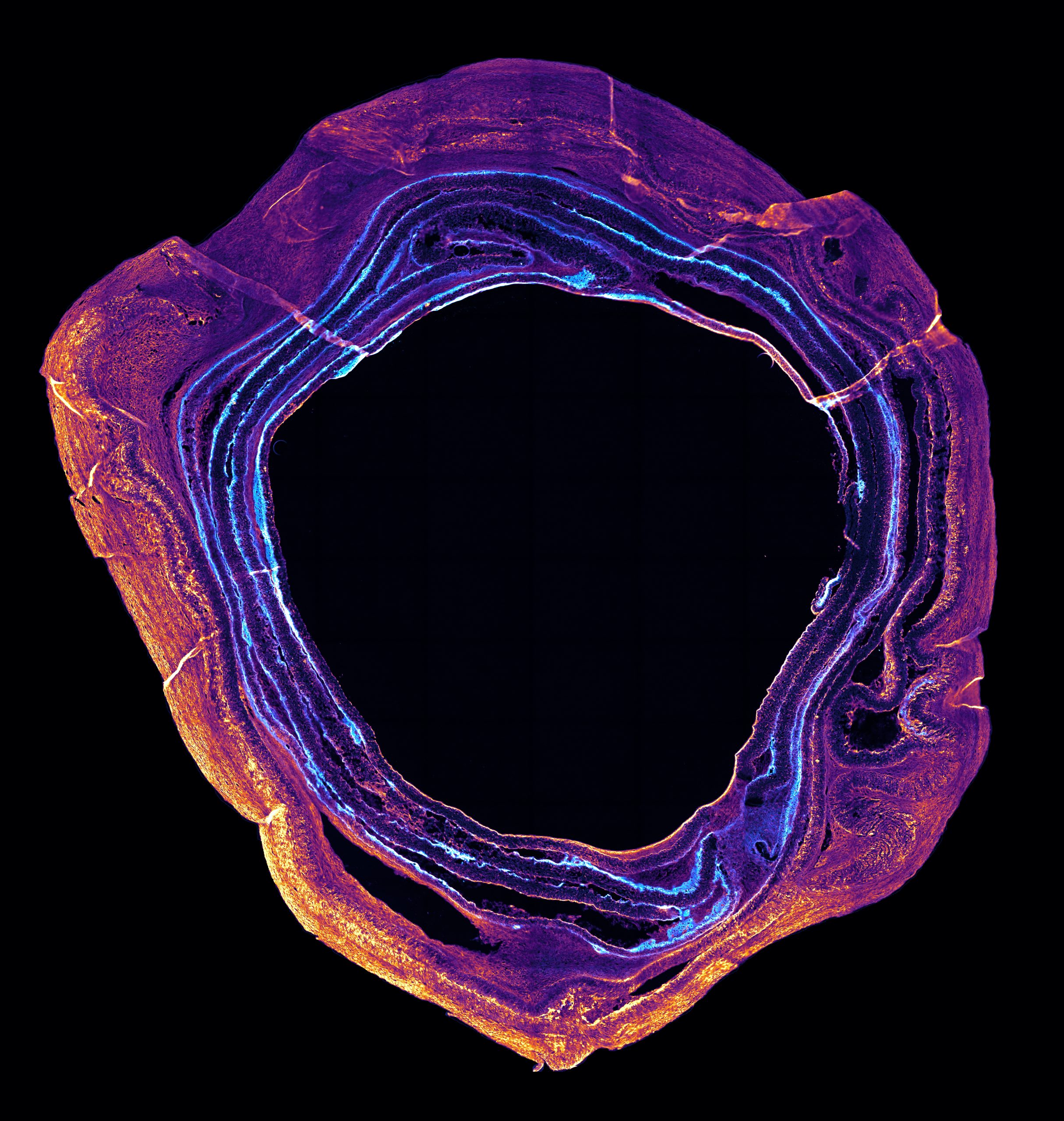
Description: The intervertebral disc (IVD) sits between the vertebrae of your spine and absorbs shock, transmits forces, and allows for a full range of motion. When the IVD degenerates, it is prone to bulging and tearing which can lead to disc herniation. That can have significant consequences, including chronic pain and even paralysis. Nearly 90% of people will develop disc degeneration throughout their lifetime making it the leading cause of lower back pain and is estimated to cost nearly 8.1 billion USD in annual Canadian health care costs. There is currently no treatment to restore native IVD function. This has led to an interest in growing an IVD in the lab that could be transplanted into patients. One of the challenges in growing the IVD in the lab is getting the different cell types to organize in a way that mimics the natural disc. My research focuses on characterizing and developing the interface between these distinct tissue types of a lab-grown disc. The inner rings of the disc are unique for their expression of type II collagen, whereas the outer rings only express type I collagen. In this image, miniature lab-grown discs follow a similar distribution of collagens.
Why did you conduct this research? During my undergrad I spent many hours in lectures learning about the most cutting-edge research and working on various research projects, the most captivating of which was the notion of growing tissues in the lab from a small pool of cells. To me, this represents the future of medical research, where one day organs can be generated from a patient’s own cells. This would resolve organ rejection, organ shortages, and long wait times on transplant lists, allowing us to live longer and healthier lives.
Technique: Intervertebral disc cells were isolated from cow tails and grown on a roll of biodegradable plastic that mimics the structure of the intervertebral disc. The miniature discs (roughly the thickness of your pinky finger) were collected after 3 weeks of growth and sliced cross-sectionally at 7 microns (approximately 1/10th the thickness of a human hair). These thin slices of the miniature discs were stained for type I (magenta/yellow) and type II (cyan/white) collagens. When viewed with a special microscope we are able to visualize and distinguish type I and II collagen within the tissue.
Acknowledgements: I would like to acknowledge the members of the Kandel lab and the Lunenfeld-Tanenbaum Research Institute Microscopy Facility for their support on my project and the collection of this image.

