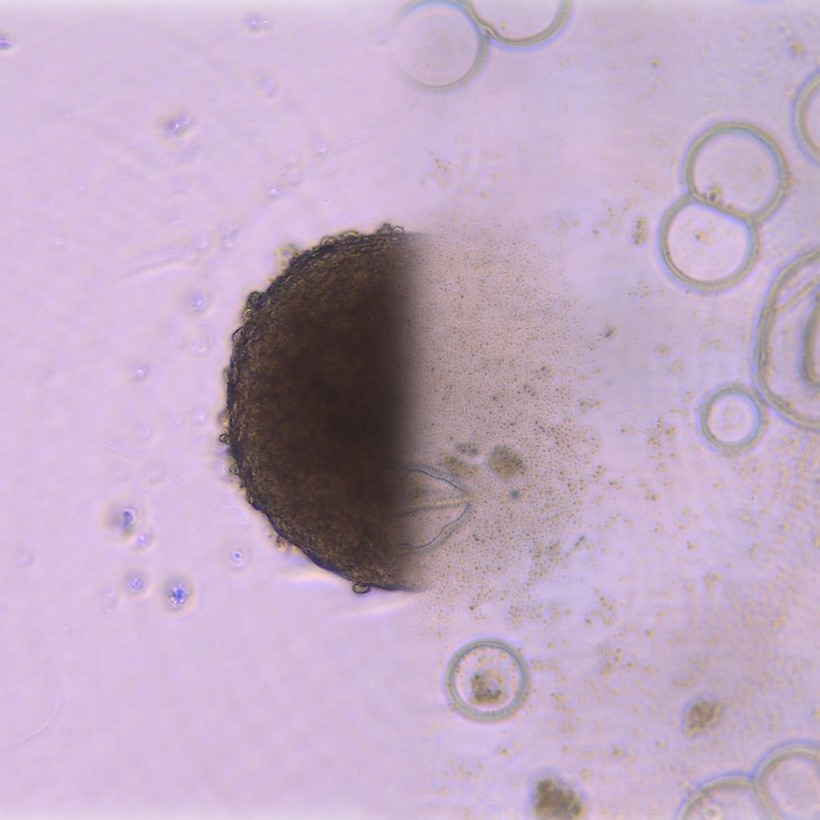
Description: The current cancer therapies lack specificity for their targets, thereby killing both healthy cells and cancer cells. This makes cancer treatments a physically and mentally traumatic experience for the patients. One of the most visible and well-known outcomes of unspecific treatments is the hair loss during chemotherapy—since chemotherapy targets all cells that rapidly divide like cancer cells do, hair follicles are also destroyed as a side-effect. Recent advancements in molecular biology had enabled the identification of molecules exclusively found in cancer cells, unveiling the potential for much more specific and effective treatments. One of these novel molecules for Head and Neck Cancers is HNCM1.
And then there were none: the image shows the head and neck cancer cells treated with the drug that targets HNCM1, which have died during the treatment. This is one of the many examples that reflect on the promising outlook of cancer treatments that will greatly enhance the cancer patients’ quality of life.
Why did you conduct this research? A downfall of longevity is the appearance of several diseases that are often associated with old age, such as cancers. One of the reasons why cancer makes such an interesting but difficult topic to investigate is the unique underlying mechanisms of how each cancer type is formed. I have conducted this research in the hopes to contribute to meeting the growing demands for targeted cancer therapy. One day, we could even witness the defeat of cancer with a single dose of “vaccine”—who knows?
Technique: The 3D cell cultures (spheroids) are created by seeding the cells to an ultra-nonadhesive surface with a round well bottom, which causes the cells to aggregate onto themselves to form a sphere. The left half of the image is a spheroid before the treatment against HNCM1, and the right half of the image is the same spheroid after 3 weeks of treatment, taken with a camera installed in the microscope. The image is modified with photoshop to adjust the contrast and the saturation of the background, as well as to merge the two images together with a fading effect.
Acknowledgements: I would like to acknowledge Drs. Thomas Kislinger Salvador Mejia-Guerrero and at the Department of Medical Biophysics for their insightful supervision throughout this project.

