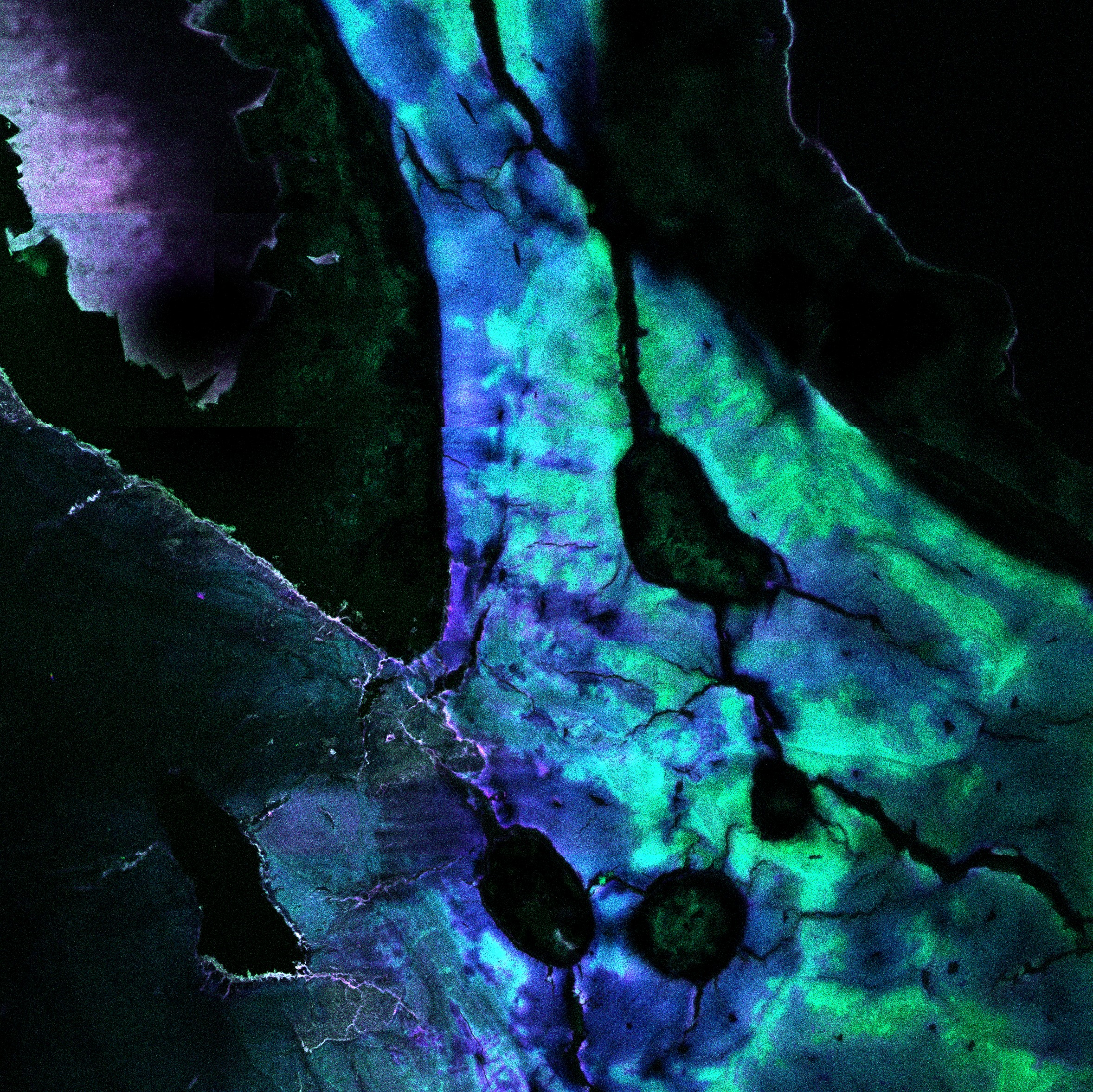
Description: This research began as a means of testing various modes of using laser scanning confocal microscopy (LSCM) on bone, particularly using various stains. I wanted to examine what would be the best method to detect histological changes in bone, particularly this difference between perimortem trauma and early postmortem damage. First, I needed to test for diagenetic changes, or postmortem histological alterations, in bone. I also needed to find out what stains would work best with the LSCM. As a means to test this, I used 3rd-5th BCE aged rib fragments stained in toluidine blue. A colleague was using these older bones for her research and we wanted to see what they would look like using LSCM. When looking at the image, the blue and purple areas are the unaltered bone, while the turquoise areas are the postmortem altered bone.
Why did you conduct this research? I conducted this research in an effort to push the boundaries of laser scanning confocal microscopy (LSCM) and bone as very little research has been conducted using this mode of histology. Originally the project was used to determine the effects of toluidine blue on bone using LSCM as a means of distinguishing between perimortem damage, but when examining the first slide of older bone, I noticed the postmortem alterations were in a turquoise colour rather than a blue/purple like all of the other slides. I tested this again with several other postmortem altered bone and found that the chemical changes all occurred in the alterations.
Technique: The image was taken using a Carl Ziess LSM880 Laser Scanning Confocal Microscope with bone stained using toludine blue.
Acknowledgements: Dr. Tracy Rogers, Ph.D. & Lelia Watamaniuk

