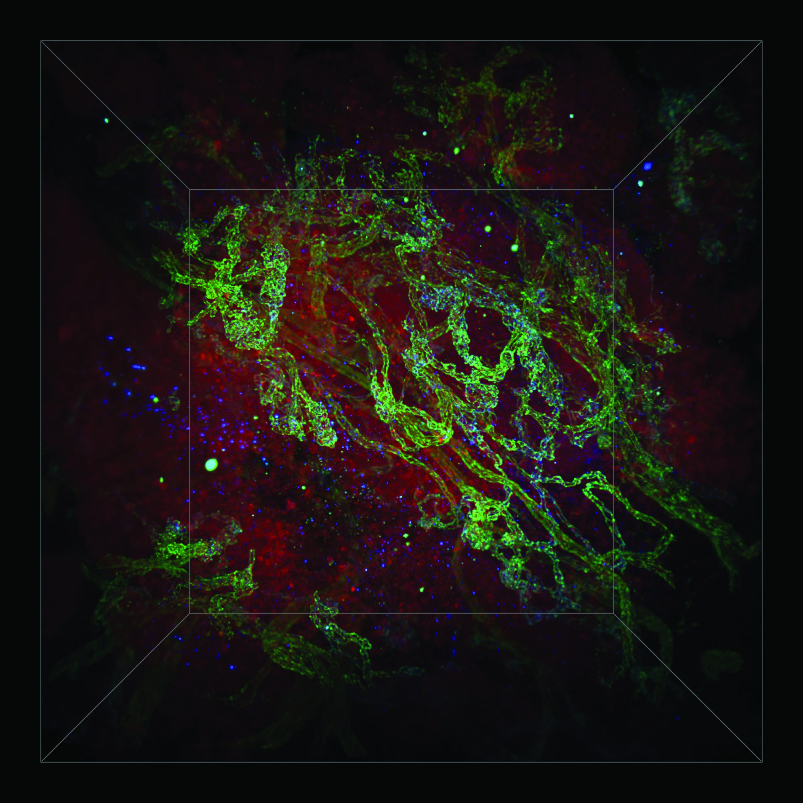
Description: Deciding on the best treatment for a patient with cancer requires a detailed profile of their tumour. We are working to develop methods that allow the different structures of the tumour to be imaged in stunning detail. State of the art technology allows researchers in our lab to visualize a patients tumour in three-dimensions with sub-cellular resolution. In this image we are viewing an ovarian tumour biopsy. This technique reveals a cosmic landscape of twisting blood vessels (blue and green) surrounded by clusters of individual tumour cells (red). The information extracted from these images forms a detailed map that is unique to each tumour. By creating detailed tumour maps scientists hope to engineer drugs that can navigate to cancer cells and eliminate them more effectively.
Why did you conduct this research? I do this research because I want to create personalized cancer treatments that are safer and more effective. I have witnessed the effect cancer has on family, friends, colleagues and the broader community. When I see people in need of better treatments I want to help. I enjoy the challenge of developing new imaging tools that allow me to visualize all the unique cells and structures that make up a tumour. I hope that by sharing images of my research people can see some beauty in a disease that is so destructive.
Technique: A three-dimensional florescent micrograph captured with a light sheet microscope of an ovarian tumour biopsy processed with tissue clearing chemicals and treated with fluorescent labels for cell nuclei (red), and blood vessel proteins (VE-Cadherin – green; PVLAP – blue).
Acknowledgements: B.R.K. would like to acknowledge the Natural Science and Engineering Research Council of Canada (NSERC), the Canadian Institutes of Health Research (CIHR), the Royal Bank of Canada, Borealis AI, the Wildcat Foundation, the Yip Family and the Dorrington Family for funding and student fellowships.

